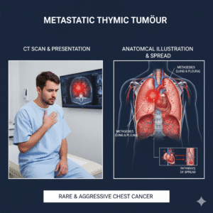
4D CT scan in Oncology
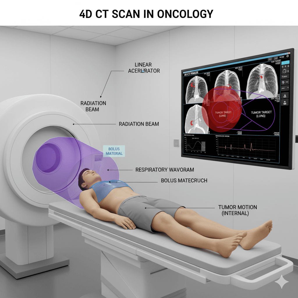
4D CT Imaging in Radiotherapy Planning
Q1: What is 4D CT Imaging?
A: 4D CT imaging is an advanced type of computed tomography (CT) scan used in radiotherapy planning. It creates detailed images of the body that show how a tumor and surrounding organs move over time, typically during the breathing cycle. The “fourth dimension” is time, allowing doctors to see not just the position of the tumor, but how it changes with motion.
Q2: How is 4D CT Imaging different from regular CT scans?
A: A regular CT scan provides a static, three-dimensional image of the body at a single point in time. In contrast, 4D CT imaging captures multiple images over time, creating a dynamic view that shows how the tumor and nearby tissues move, particularly with breathing. This added time dimension is crucial for planning precise radiation treatments, especially for tumors in areas like the lungs, abdomen, and chest, where movement can affect accuracy.
Q3: Why is 4D CT Imaging important in radiotherapy planning?
A: 4D CT imaging is important because it helps radiation oncologists understand how a tumor moves and changes shape during the breathing cycle. This information allows them to:
- Precisely target the tumor: By accounting for movement, doctors can better direct radiation at the tumor while sparing healthy tissues.
- Reduce side effects: With a clearer understanding of tumor motion, radiation can be delivered more accurately, minimizing damage to surrounding healthy tissue.
- Improve treatment effectiveness: Accurate targeting ensures the tumor receives the full intended dose of radiation, which can improve treatment outcomes.
Q4: How is 4D CT Imaging used in the radiotherapy process?
A: 4D CT imaging is typically used during the planning phase of radiotherapy. The process involves:
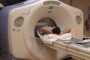
- Scanning: The patient undergoes a 4D CT scan, during which images are taken throughout the breathing cycle.
- Analyzing motion: The resulting images are analyzed to track how the tumor and nearby organs move over time.
- Planning treatment: Based on this information, radiation oncologists create a treatment plan that takes tumor motion into account, ensuring that the radiation targets the tumor accurately, even as it moves.
Q5: Who benefits from 4D CT Imaging?
A: 4D CT imaging is particularly beneficial for patients with tumors that move during normal bodily functions, such as:
- Lung cancer: Where tumors can move significantly with each breath.
- Liver cancer: As the liver moves with the diaphragm during breathing.
- Breast cancer: To avoid unnecessary radiation to the heart and lungs.
Patients with tumors in other areas of the body that are affected by movement may also benefit from 4D CT imaging.
Q6: Is 4D CT Imaging safe?
A: Yes, 4D CT imaging is safe. It involves a similar process to a regular CT scan but captures images over a longer period. While it does involve exposure to radiation, the benefits of more precise radiotherapy planning typically outweigh the risks. Your healthcare team will ensure that the radiation dose is as low as possible while still providing the necessary information.
Q7 What are the different methods used in 4D CT Imaging?
A: Several methods are used to capture and account for tumor movement in 4D CT imaging. The most common methods include:
- Varian RPM (Respiratory Gating System)
- Phase-Based Imaging
- Amplitude-Based Imaging
- Surrogate Marker Systems
Varian RPM System
A: The Varian Real-time Position Management (RPM) system is a respiratory gating system used during 4D CT imaging and radiotherapy. It tracks the patient’s breathing in real time using an infrared camera and a reflective marker placed on the patient’s chest or abdomen. The system monitors the movement of the marker as the patient breathes, synchronizing the CT scan or radiation delivery with specific points in the breathing cycle. This ensures that the tumor is in the correct position for accurate imaging and treatment.
Phase-Based Imaging in 4D CT?
A: Phase-based imaging divides the breathing cycle into different phases, such as inhalation, exhalation, and the transitions between them. During the 4D CT scan, images are captured at each phase of the breathing cycle. This method helps create a comprehensive picture of how the tumor moves throughout the entire cycle, allowing for precise targeting of radiation at the most stable phases.
Amplitude-Based Imaging
A: Amplitude-based imaging focuses on the extent of movement (amplitude) during the breathing cycle rather than specific phases. It records the position of the tumor when it reaches certain amplitudes of motion, such as when the tumor is at its highest or lowest point in the chest. This method is useful for capturing the full range of tumor motion and helps in planning radiation delivery when the tumor is within a specific range of motion.
Surrogate Marker Systems
A: Surrogate marker systems use external markers or devices placed on the patient’s body to track respiratory motion. These markers move with the patient’s breathing and provide a real-time reference for the position of the tumor. This information is then correlated with the CT images to map out tumor motion accurately. Surrogate markers can be non-invasive and are often used in combination with other respiratory gating techniques.
Q8: Why are these methods important in radiotherapy planning?
A: These methods are crucial because they help radiation oncologists account for the natural movement of tumors during breathing. By understanding how a tumor moves, doctors can plan radiotherapy that precisely targets the tumor while mini
Q9: How should I prepare for a 4D CT Scan?
A: Preparation for a 4D CT scan is usually straightforward. You may be asked to wear comfortable clothing and remove any metal objects, such as jewelry. In some cases, you might be given specific breathing instructions to help capture clear images. Your healthcare team will provide detailed instructions based on your specific situation.
Q10: Where can I receive 4D CT Imaging for radiotherapy planning?
A: 4D CT imaging is available at cancer treatment centers and hospitals with advanced radiotherapy facilities. If your treatment plan would benefit from 4D CT imaging, your radiation oncologist will refer you to a center that offers this technology.
If you have more questions about 4D CT imaging or how it might be used in your treatment, feel free to contact us for guidance on how this technology can help improve the accuracy and effectiveness of your radiotherapy.
Related Post

CyberKnife
August 6, 2024

Immunotherapy
August 7, 2024
MRI Linac
August 7, 2024

Gamma Knife
August 7, 2024
Cancer Screening
August 22, 2024
Gallery
Click below to book a clinic appointment
Ask More Questions Send Query On Email


